
- 79
- 1 203 450
Radquarters
Приєднався 13 бер 2016
Let's learn some radiology! Here you'll find high-yield, educational radiology lectures with an emphasis on body imaging using a multimodality approach, including MRI, CT, ultrasound, radiography, and nuclear medicine. These video lectures are designed for radiology residents, fellows and imaging technologists, as well as any student or practitioner interested in optimizing patient care through radiology.
The information on this channel is for educational purposes only, and is not intended or implied to be a substitute for professional medical advice, diagnosis or treatment.
- Daniel Kowal, MD, RMSK
Board certified, fellowship trained radiologist practicing in Massachusetts.
The information on this channel is for educational purposes only, and is not intended or implied to be a substitute for professional medical advice, diagnosis or treatment.
- Daniel Kowal, MD, RMSK
Board certified, fellowship trained radiologist practicing in Massachusetts.
Ultrasound of Interstitial Ectopic Pregnancy
In this radiology lecture, we review the ultrasound appearance of interstitial ectopic pregnancy!
Key teaching points include:
1) Interstitial ectopic pregnancies are rare, occurring in proximal (interstitial) portion of fallopian tube within muscle wall of uterus
2) Much less common than tubal ectopic pregnancy occurring in the more distal ampullary and isthmic portions of fallopian tube
3) Interstitial ectopic pregnancies are important because higher morbidity and mortality due to risk of life-threatening hemorrhage
4) Abnormally eccentric gestational sac with thin surrounding myometrium: less than 5 mm myometrial thickness highly suspicious
5) “Interstitial line” sign: Thin echogenic line extending from endometrial cavity to ectopic gestational sac. Thought to represent interstitial portion of tube separating the ectopic pregnancy from the endometrium
6) Medical: Systemic MTX, may also be injected into sac
7) Surgery: Cornual wedge resection when ruptured versus hysterectomy
8) Can be confused with angular pregnancy: Rare, intrauterine pregnancy with implantation eccentrically high at the lateral angle of uterine cavity. More medial than interstitial ectopic pregnancies. No interstitial line sign, and greater than 5 mm thickness of overlying myometrial mantle
9) Angular pregnancy can result in normal pregnancy, but increased risk of miscarriage and uterine rupture. Should be followed closely to ensure growth towards endometrial cavity
10) Angular pregnancy is sometimes referred to as a “cornual pregnancy,” but controversial as earliest use of term cornual pregnancy refers to intrauterine implantations in anomalous unicornuate, bicornuate or septate uteri. To avoid confusion, best to specifically describe whether the gestational sac is intrauterine or ectopic
To learn more about the Samsung RS85 Prestige ultrasound system, please visit: www.bostonimaging.com/rs85-prestige-ultrasound-system-4
Click the UA-cam Community tab or follow on social media for bonus teaching material posted throughout the week!
Website: Radquarters.com/
Spotify Video Podcast: spoti.fi/462r0F2
Apple Video Podcast: apple.co/3ZhHuGu
Instagram: Radquarters
Facebook: Radquarters/
X (Twitter): Radquarters
Reddit: www.reddit.com/user/radiologistHQ/
Переглядів: 8 509
Відео


Ultrasound of Ovarian Serous Cystadenocarcinoma
Переглядів 9 тис.2 місяці тому
In this radiology lecture, we review the ultrasound appearance of ovarian serous cystadenocarcinoma! Key teaching points include: 1) Serous cystadenocarcinoma is the common ovarian malignancy and most common ovarian epithelial tumor 2) High-grade and low-grade types 3) Peak incidence 6th-7th decades 4) Ultrasound appearance: Mixed cystic and solid mass with papillary projections and thick septa...

Ultrasound of Parathyroid Adenoma
Переглядів 9 тис.2 місяці тому
In this radiology lecture, we review the ultrasound appearance of parathyroid adenoma! Key teaching points include: 1) Benign tumor of the parathyroid glands 2) Most common cause of primary hyperparathyroidism: Elevated serum calcium and parathyroid hormone (PTH) levels 3) Ultrasound: Solid, homogeneous and very hypoechoic. Oval or bean-shaped, long axis oriented craniocaudal. Hypervascular. Ma...

Ultrasound of Parotitis
Переглядів 9 тис.3 місяці тому
In this radiology lecture, we review the ultrasound appearance of parotitis in the pediatric population! Key teaching points include: 1) Parotitis = Inflammation of the parotid glands 2) Acute parotitis is usually infectious, most commonly viral 3) Mumps is most common viral cause in children, often bilateral 4) Bacterial parotitis can cause suppurative parotitis seen in premature infants and i...

Ultrasound of Sublingual Dermoid Cyst
Переглядів 9 тис.4 місяці тому
In this radiology lecture, we review the ultrasound appearance of sublingual dermoid cyst and explain floor of mouth anatomy! Key teaching points include: 1) The floor of the mouth is a horseshoe-shaped area beneath tongue and in between sides of mandible, inferiorly bounded by mylohyoid muscle, and containing sublingual space (SLS) 2) SLS medial border: Midline genioglossus/geniohyoid muscle c...
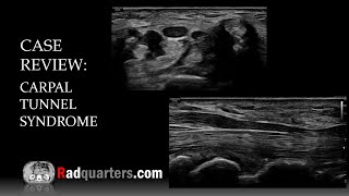
Ultrasound of Carpal Tunnel Syndrome
Переглядів 14 тис.8 місяців тому
In this radiology lecture, we review the ultrasound appearance of carpal tunnel syndrome! Key teaching points include: 1) Carpal tunnel syndrome results from median nerve compression and is the most common upper extremity entrapment neuropathy. 2) With carpal tunnel syndrome, see hypoechoic enlargement of the median nerve as enters carpal tunnel with flattening of nerve = Notch sign, also volar...

Ultrasound of Ganglion Cyst & Wrist Anatomy Review
Переглядів 8 тис.9 місяців тому
In this radiology lecture, we review the ultrasound appearance of ganglion cysts while highlighting relevant wrist ultrasound anatomy! Key teaching points include: 1) Ganglion cysts are viscous, mucin-filled collections lacking a synovial lining 2) Most commonly occur at hand/wrist = Most common wrist mass 3) Location: Dorsum of wrist (60%), frequently adjacent to scapholunate ligament; volar w...

Radquarters update
Переглядів 6899 місяців тому
Radiologist Headquarters has a new name: Radquarters! Same high-yield content, but now with a streamlined name that's easier to remember. Click the UA-cam Community tab or follow on social media for bonus teaching material posted throughout the week! Website: Radquarters.com/ Spotify Video Podcast: spoti.fi/462r0F2 Apple Video Podcast: apple.co/3ZhHuGu Instagram: Radquarters Face...

Ultrasound of Epididymitis & Orchitis
Переглядів 19 тис.10 місяців тому
In this radiology lecture, we review the ultrasound appearance of acute epididymitis and orchitis! Key teaching points include: 1) Epididymitis = Inflammation of epididymis. Usually bacterial, most commonly due to retrograde ascent from bladder or prostate. 2) Causative infectious agent varies based on age: Adults younger than 35: Neisseria gonorrhoeae, Chlamydia trachomatis (STDs). Adults olde...
Ultrasound of Acute Cholecystitis
Переглядів 12 тис.11 місяців тому
In this radiology lecture, we review the ultrasound appearance of acute cholecystitis, including gangrenous and emphysematous cholecystitis! Key teaching points include: 1) Acute cholecystitis = Acute gallbladder inflammation. 2) Most often (95%) caused by an impacted, obstructing gallstone in the cystic duct or gallbladder neck = Acute calculous cholecystitis. 3) Clinically presents as persist...
Ultrasound of Intussusception
Переглядів 19 тис.Рік тому
In this radiology lecture, we review the ultrasound appearance of ileocolic and small bowel-small bowel intussusception in children! Key teaching points include: 1) Intussusception occurs when bowel is pulled into itself or into neighboring bowel. 2) Intussusceptum is the prolapsing bowel pulled into intussuscipiens which receives the bowel. 3) Two major types: Ileocolic and small bowel-small b...
Ultrasound of Polycystic Ovarian Syndrome
Переглядів 22 тис.Рік тому
In this radiology lecture, we review the ultrasound appearance of polycystic ovarian syndrome (PCOS)! Key teaching points include: 1) PCOS often presents with the clinical triad of oligomenorrhea and/or anovulation, hirsutism, and obesity. Associated with subfertility and recurrent pregnancy loss. 2) Rotterdam criteria (2003) states that PCOS diagnosis requires at least two of the following: Ol...
Ultrasound of Pleomorphic Adenoma of the Parotid Gland
Переглядів 10 тис.Рік тому
In this radiology lecture, we review the ultrasound appearance of pleomorphic adenoma of the parotid gland! Key teaching points include: 1) Pleomorphic adenoma AKA benign mixed tumor. 2) Most common salivary gland tumor, most common benign salivary gland tumor, and most common in the parotid gland. 3) Most common in patients aged 40-50, slightly more common in females. 4) For salivary gland mas...
Ultrasound of Epidermal Inclusion Cyst
Переглядів 8 тис.Рік тому
In this radiology lecture, we review the ultrasound appearance of epidermal inclusion cyst! Key teaching points include: 1) Epidermal inclusion cyst is the most common cutaneous cyst. 2) Can occur anywhere: Head, neck, trunk, extremities. 3) Benign, keratin-containing cyst lined by a wall of stratified squamous epithelium. 4) On ultrasound, appears as a well-circumscribed, round to oval mass wi...
Ultrasound of Torsion of the Appendix Testis
Переглядів 8 тис.Рік тому
In this radiology lecture, we review the ultrasound appearance of torsion of the appendix testis and appendix epididymis! Key teaching points include: 1) Appendix testis is a vestigial appendage usually located between upper pole of testis and head of epididymis. 2) AKA hydatid of Morgagni, the appendix testis is commonly present as a normal finding. 3) Appendix epididymis typically arises from...
Hello sir, how much volume of contrast and flow rate should be followed in hepatic protocol
Excellent presentation. If possible a video could be made comparing Hepatic Abcess and Hydatid Disease.
Super.thank you
You're welcome, glad you enjoyed it!
👏👏👏👏👏👏👏👍👍👍👍👌👌👌👌
Bravo Professor
Thanks!
Bravo Professor
Thank you!
Briliant, many thanks!
Glad you enjoyed it!
Briliant style and love the quick review at the end!
Thank you!
Beautiful! Amazing how clearly the 3D Imaging delineates the location of the gestational sac as compared to 2D in this case.
Agreed, the 3D is very helpful!
thanks sir 😊
You are most welcome!
Great one! Thanks
Welcome!
This was helpful and clear. Thank you!
Great to hear, and thanks for watching!
Thank you for sharing. Always so educational.
Thank you, appreciate that!
@@Radquarters would you suggest any resources for obstetrics and gynecology scans?
@@johnweak3198 Ultrasound: The Requisites is an excellent book, and one I recommend for my students.
@@Radquarters thank you very much!
very nice
Thank you! Appreciate that
Ultrasound while lying can identify varicocele?
Yes, particularly if Valsalva is used, but standing position often helps with diagnosis.
Very nice ultrasound .which model used
Thanks! These images were obtained on a Samsung RS85 Prestige unit: www.bostonimaging.com/rs85-prestige-ultrasound-system-4
I get it that you are really smart and stuff but it takes a real smart person to be able to dumb it down so regular people can understand what the f*** you're talking about
Can we do it in ge 16 slice machine.. we don't get this kind of scan ..our del ded is 8
Yes, a multiphase liver scan can be performed with a 16 slice scanner.
@@Radquarters yes sir but we r not getting proper arterial phase our scan del sec is 7 to 8 sec
@@jeevnasam4810 You might want to consider a delay closer to 35 seconds for the late hepatic arterial phase. 7-8 seconds is typically closer to a true arterial phase (i.e., hepatic artery enhancement with no portal venous enhancement).
Great presentation.. thanks from India.
Glad you liked it, and hello to India!
Great work! 😀
Thank you!
Great video. Thank you, and keep up the excellent work. I appreciate it 😀
Thank you for the kind feedback!🙂
Thank you!
You're welcome!
This presentation was really good. Well done and keep up the good work. Thank you for your efforts.
Thank you for the feedback, and I'm glad you enjoyed it!
Desperate Housewives S2 E12 brought me here. LOL
Ha! Thanks for visiting :)
Doctor, after take a medicine i feel better, but today i feel the pain again after 3 week the pain is gone... Is it normal??
Though its educational , but you speak too fast with monotonous voice.
Thank you, very informative
Glad you enjoyed it!
Áreas sólidas no grasas , volumen probablemente sobre 500 CC, vascularizadas. GIRADS 5. Sobre el 90% riesgo de Cáncer
👏👏
Thank you!
💥👏🤲
Thanks!
Good case
Thanks!
@@Radquarters sir I am radiology resident Kindly do more 5 minute usg cases .Those cases were tooo good
O´TIMOS CASOS
Thank you!
Great and illuminating presentation , Dr Daniel!
Thank you, and so glad you enjoyed it!
Beatutiful film! May i know frequency working probe? thanks
Thank you! For wrist ultrasounds, I tend to use a combination of a 2-14 MHz and a 4-18 MHz linear transducers. A hockey stick (3-22 MHz) can also be helpful for more focused evaluation (ligaments, nerves).
@@Radquarters thank you so much
Great👏👏👍👍
Thank you!
Thanks for knowledge on it
Sure thing
Hi Should testes ultrasound be conducted with the patient standing or supine? I had a physical examination and my GP suspects varicocele, but my testes ultrasound showed no varicocele.
Scrotal ultrasounds are usually performed in supine position, but for varicocele the standing position is sometimes added.
Superb presentation 🎉
Thanks so much!
Hello ...dose this infection epididymitis orchitis damage the microtubes from the epididymis? it seems nobody talks about this posible trauma after the infection is over
Good question! Tubular ectasia of the rete testis can occur in the post-inflammatory setting. Also, depending on the severity and duration of epididymo-orchitis, both the epididymis and testis can develop heterogeneous parenchyma +/- calcifications.
My situation right now..am so stressed
Amazing video!
Thanks Talia Cheng, appreciate that!
Thanks. Very nice video.
Thank you, appreciate that :)
Very good example! Although I think you should explain that parathyroids can be located in many places including intrathyroid, and because of that we can speak about parathyroid change in the thyroid. Also lack of TiRADS which should be 4 in this case. A plus for typing what US system you use, a top one.
Thank you, and excellent points! However, I do actually describe the ectopic locations of parathyroids at time 3:58 in the video, and also that they can mimic TI-RADS 4 nodules at 4:14.
@@Radquarters I missed it somehow, my fault, forgive me, do you perform echocardiography as well?
@@adamguszak7050 No problem, and thanks for watching. I don't perform echocardiography. Sorry!
thank youuuuu from algeria
You're welcome, and great to hear from Algeria!
Thank you❤
Great emergency cases
Thanks, and glad you enjoyed them!
High quality images, Can you tell me the Program or application you use to Record your nice videos?
Sure, I make the presentations using PowerPoint, then I record and edit using ScreenPal. I also use a Blue Yeti mic.